PINEAL GLAND Disrupted by Heavy Metals and Glyphosate
PINEAL GLAND Disrupted by Heavy Metals and Glyphosate
Sulfate, Sleep and Sunlight: The Disruptive and Destructive Effects of Heavy Metals and Glyphosate
By Claire I. Viadro, MPH, PhD
Neurological disorders, autoimmune diseases—they seem to be everywhere these days. Scientists writing in Neurology in 2007 estimated that the burden of neurologic illness affects “many millions of people in the United States.”1
Autoimmune illness, too, is at epidemic proportions—nearly 24 million Americans as of 2012.2 These trends are disturbing enough in their own right, but even more disturbing is the general scientific apathy about why the surge in these diseases is occurring.
Why do the causes of these alarming epidemics remain “underrecognized and underaddressed?”3
Stephanie Seneff is one of the all-too-rare scientists who is trying to ask the questions and connect the dots. Dr. Seneff4
is a senior research scientist at the MIT Computer Science and
Artificial Intelligence Laboratory with an illustrious career and
lengthy publication record.
Of late, she has been using computer science and natural language
processing (NLP) techniques (NLP is a field of computer science,
artificial intelligence, and linguistics) to delve into the impact of
environmental toxins on human health.
She has developed some particularly convincing hypotheses relating to
autism and, more recently, cancer. At the Third International Symposium
on Vaccines,5
presented in March 2014 as part of the 9th International Congress on
Autoimmunity, Dr. Seneff was one of 15 speakers invited to present
scientific research by the Children’s Medical Safety Research Institute6 (CMSRI) on the adverse health effects of aluminum adjuvants and aluminum-adjuvented vaccines.
She discussed “a role for the pineal gland in neurological damage
following aluminum-adjuvented vaccination.” Along the way, she made many
fascinating connections between various strands of her recent work,
briefly summarized in this article.
The Critical Role of Sulfate
Dr. Seneff persuasively makes the case that neurological
brain diseases have a common origin that begins with an insufficient
supply of sulfate to the brain. Sulfate is the oxidized form of sulfur.
Dr. Seneff has argued that systemic sulfate deficiency “may be the most
important factor in many of the health issues facing us today.”7
I’ll get to her thoughts on why so many people are deficient in
sulfate in a moment, but suffice it to say that one of the consequences
of insufficient sulfate in the brain is that it impairs the brain’s
ability to eliminate heavy metals and other toxins. To make matters
worse, those same toxic metals also interfere with sulfate synthesis.
The net result can be an accumulation of cellular debris.
How do our brains get rid of cellular debris? Dr. Seneff cited recent work showing that sleep is crucial in this regard.8 Sleep is the brain’s “housekeeper.”
This housekeeping takes place in the lysosomes. (Lysosomes—the cell’s
waste disposal system—are filled with enzymes that break down unwanted
materials.) However, the lysosomes cannot perform their important
clearing work without sulfate, specifically heparan sulfate.
Heparan sulfate belongs to the family of glycosaminoglycans or
GAGs—complex polysaccharides that provide structural integrity to cells.
Heparan sulfate regulates many different biological functions,
including ion and nutrient transport as well as molecular signaling
cascades for most of the body’s cells.
It also plays an important role in fetal brain development.9
The role of heparins and heparan sulfate as “endogenous antioxidants”
protecting against damaging free radicals was recognized over 20 years
ago.10
Interesting evidence of what happens when heparan sulfate is
deficient comes from human and mouse studies of autism. In one study,
“designer” mice engineered to have impaired heparan sulfate synthesis in
the brain displayed all the classic features of autism, including
sociocommunicative deficits and stereotypies.11
To understand this area of autism research, it is helpful to
visualize the ventricles in the brain. The ventricles—a “communicating
network of cavities filled with cerebrospinal fluid (CSF)”12 —consist of two lateral ventricles, the third ventricle, the cerebral aqueduct, and the fourth ventricle.
A 2013 study in Behavioural Brain Research found heparan
sulfate deficiency in the subventricular zone of the lateral ventricles
of four autistic individuals. The authors posited that this “may be a
biomarker for autism, and potentially involved in the etiology of the disorder“13 [emphasis added]. Other studies have identified heparan sulfate depletion in the third ventricle.14
Enter the Pineal Gland
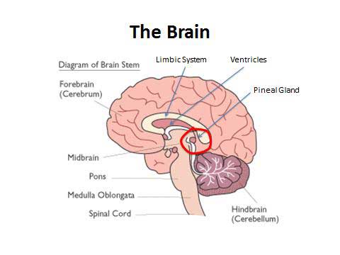
A notable feature of sulfate is that it is difficult to transport.15
Dr. Seneff’s extensive work on sulfur deficiency has led her to
consider the important but perhaps underestimated role of the pineal
gland in the transport process. The pineal gland is a neuroendocrine
organ of the brain that resides in close proximity to the ventricles, as
seen in the following illustration.
Figure 1. The brain and the pineal gland
A key role of the pineal gland is to synthesize and secrete melatonin, which controls the sleep/wake cycle.16
Dr. Seneff suggests that one of the critical purposes of melatonin, in
turn, is to deliver sulfate to the neurons at night during sleep. In her
words, melatonin is clearly a “sulfate delivery system.” Dr. Seneff
outlined this intricate and elegant delivery system as follows:
- With sunlight exposure serving as a catalyst, the pineal gland
builds up supplies of sulfate by day, storing it in heparan sulfate
molecules.
- The pineal gland produces melatonin in the evening, transporting it
as melatonin sulfate to various parts of the brain, including the third
ventricle, where the melatonin releases the sulfate into the CSF.
The association of autism with heparan sulfate depletion in the lateral and third ventricles17, 18
now gets more interesting, because the tip of the third ventricle is
encased in the pineal gland. The pineal recess is in fact the “main site
of penetration of melatonin into the CSF.”19
In other words, under normal circumstances the pineal gland delivers
melatonin sulfate to the third ventricle, which then diffuses the
sulfate throughout the CSF. In addition, melatonin not only transports
sulfate but also is an outstanding antioxidant and binds toxic metals to
help dispose of them. It may come as no surprise, then, that melatonin
impairment has been implicated in autism.
In healthy individuals, melatonin also plays an important role in
inducing REM sleep, which may be the most important stage of sleep.
Interestingly, Alzheimer’s disease is associated with reduced REM sleep
and a calcified pineal gland. Sleep disorders are also linked to autism
as well as other neurological diseases, including depression,
schizophrenia, ALS, Parkinson’s disease, and others.
Your Pineal Gland and Heavy Metals
If one recognizes that heavy metals play a part in the
modern-day epidemic of neurological diseases, then part of the
explanation for the sleep disorders encountered in various neurological
diseases may be that both aluminum and mercury (thimerosal) disrupt the
pineal gland and its ability to make sulfate. When the pineal gland’s
ability to make sulfate is impaired, this, in turn, reduces production
of melatonin, all-important for adequate and healthy sleep. The pineal
gland is particularly susceptible to aluminum and other heavy metals
because it is not protected by the blood-brain barrier and has a very
high blood perfusion rate.
The pineal gland’s vulnerability to aluminum is illustrated in a 1996
paper showing that the concentrations of aluminum in the pineal gland
were “consistently observed” and “markedly higher” than in other brain
tissues examined (pituitary, cortex, and cerebellum).20
Returning to the link between the pineal gland, heavy metals, and
sleep, a telling fact gleaned by Dr. Seneff from the national Vaccine
Adverse Event Reporting System (VAERS) is that insomnia occurs more
often as an adverse reaction to aluminum-containing vaccines than to
vaccines not containing aluminum.
Scientists are taking note of the fact that we live in an “age of
aluminum,” with aluminum exposure occurring through vaccines as well as
multiple other channels.21, 22
Moreover, although many experts would have us believe that the question
of thimerosal and vaccine safety went away after federal agencies
issued lukewarm recommendations to reduce its use as a vaccine
preservative in the early 2000s, Dr. Seneff noted that thimerosal is
still very much relevant.
Why Some Children May Be More Prone to Vaccine Damage
She called attention to a 2013 paper that reminds us that
thimerosal not only is not found in nature but is a “designer” mercury
compound created by humans that is the most toxic nonradioactive
metal—”even more toxic than lead to human fetal and neuronal cells.”23
Bringing things full circle back to sulfur/sulfate, Dr. Seneff pointed
out that the article makes an important link between autism and
sulfation, concluding that children with abnormal sulfation chemistry
(among other factors) may be particularly susceptible to the toxic
effects of the thimerosal in flu and other childhood vaccines.24
In fact, due to expanded recommendations for flu shots in pregnant
women and young children, exposure to thimerosal through vaccination has
remained widespread in the US, and more than half of all flu vaccine
doses are still thimerosal-preserved.25 Incredibly, the authors of the 2013 paper note the following:
“Estimates are that the maximum lifetime exposure to [thimerosal] a vaccinated person may receive is now more than double what it would have been had the pre-2000 vaccination schedule been maintained.”26 [Emphasis added]
Dr. Seneff has done a lot of investigations using the VAERS database,
which—despite its limitations—can be very informative. She notes that
concurrent with the aggressive peddling of thimerosal-containing flu
shots and other aluminum-containing vaccines, there has been a rise in
reporting of both vaccine adverse events and autism spectrum disorders.
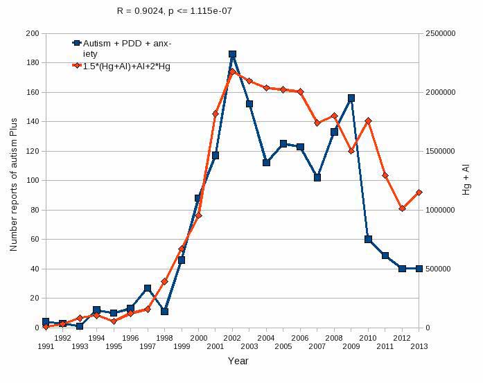
She described one careful analysis of the VAERS database.
In a graph that speaks for itself (Figure 2 below), she plotted the
number of VAERS reports mentioning three types of adverse events
(autism, pervasive developmental disorders or PDDs, and anxiety
disorder) against the total burden of two heavy metals (aluminum and
mercury) in vaccines according to the current vaccine schedule. One can
immediately see that the adverse event and heavy metals lines are quite
similar. Moreover, both lines show a sharp spike around the year 2000,
which is when the burden of aluminum and thimerosal increased. Dr.
Seneff commented that while aluminum and thimerosal are each bad enough
on their own, they also work synergistically to cause harm.
Figure 2. Link between reports of vaccine adverse events (VAERS
database) and aluminum (Al) and mercury (Hg) burden in current vaccine
schedule (Figure kindly provided by Nancy Swanson)
The Role of Sunlight
Returning once again to the topic of sulfate, Dr. Seneff
underscored the important and neglected fact that sunlight is absolutely
essential for human health because of its role in catalyzing sulfate
production. We will be sulfate-deficient if we do not get enough sun.
The pineal gland plays an important role in this process. Specifically,
endothelial and neuronal nitric oxide synthase—both of which are present
in the pineal gland—produce sulfate from reduced sulfur sources
catalyzed by sunlight.
When this process is impaired through lack of sunlight exposure, the
result is sulfate deficiency and—where a serum sulfate deficiency is
present—an individual will also have an impaired ability to dispose of
aluminum.27
Aluminum accumulation in the pineal gland over time will disrupt
sulfate supplies to the brain by interfering with the pineal gland’s
ability to make sulfate. High-SPF sunscreens are one way in which the
body can accumulate not-insignificant amounts of aluminum through skin
absorption. Sunscreens contain aluminum nanoparticles, which are more
dangerous than larger-sized aluminum particles and highly destructive in
the brain.
A 2012 study found that nanoalumina destroyed mitochondria (thus
severely depleting ATP, the body’s energy source), induced autophagy and
programmed cell death in brain endothelial cells, and decreased
expression of tight-junction proteins, thereby contributing to elevated
blood-brain barrier permeability.28
The nanoparticle effects were persistent and damaging. Thus, contrary
to popular opinion, use of sunscreen is neither beneficial nor safe.
(Dr. Seneff noted in passing that wearing sunglasses is also a terrible
idea.)
Sunlight May Be Protective Against Autism
Dr. Seneff further assessed the importance of sunlight by compiling data from demographic studies in the 50 states (Table 1).
Table 1. Correlation of sunlight exposure and autism in public school students in 50 states (grades 1–6, 2007–2008)
| Demographic |
Pearson Correlation Coefficient |
Category |
| Number of clear days |
-0.40 |
Sunlight exposure |
| Rainfall and latitude |
+0.34 |
Sunlight exposure |
| Vaccination rate |
+0.38 |
Aluminum, mercury |
Public schools in the US keep track of the number of
students enrolled in each grade, and they also keep track of the number
of students enrolled in programs specifically targeting autism. Using a
ratio of these numbers, Dr. Seneff and co-investigators calculated a
measure for autism in each state (using grades one to six for the
2007-2008 school year). They also obtained data for weather-related
factors, using these as proxies for sun exposure (e.g., number of clear
days and a combination variable capturing latitude and rainfall) and
looked at states’ vaccination rates as a proxy measure for aluminum and
thimerosal exposure.
They then calculated Pearson’s correlation coefficients as a way of
understanding the strength of the relationship (or correlation) between
sunlight exposure and autism. (Correlation coefficients range from -1.0
to 1.0, and a coefficient that is close to zero signals a weak
relationship.) Bearing in mind that correlation does not necessarily
mean causation, their analysis nonetheless produced correlations
suggesting that sunlight is protective against autism, although other
factors also clearly explain some of the variability.
One of the ways that the protective effect of sunlight exposure makes
sense is recognizing the critical role that vitamin D plays in sulfate
homeostasis. A study in mice found that activated vitamin D prevented
sulfate wasting from the kidney in urine, and mice engineered to have
defective vitamin D receptors (or with vitamin D deficiency) had
significantly reduced serum sulfate levels, which were associated with
sulfate depletion in the skeleton. Children with autism have high
sulfate in their urine but low serum sulfate levels, which clearly
indicates both generic sulfate deficiency and vitamin D deficiency.
Glyphosate: The Elephant in the Room
Dr. Seneff began paying attention to glyphosate after she
had been intensely researching autism for five or six years. Glyphosate
is a broad-spectrum systemic herbicide (known to the world under its
trade name Roundup®). Among its many nefarious health effects,
glyphosate disrupts the way the body manages sulfur.
In the process of examining all the known toxic chemicals in the
environment and assessing which one(s) would be most likely to be causal
for autism—given the specific comorbidities associated with autism—Dr.
Seneff found that glyphosate matched up almost perfectly.
Both glyphosate and autism are associated with low melatonin,
impaired sulfur metabolism (and low serum sulfate), low vitamin D, sleep
disorders, disrupted gut bacteria, and more. Glyphosate—already a very
dangerous chemical on its own—causes aluminum to be much more toxic.
Glyphosate and aluminum can be viewed as “partners in crime,” working
synergistically with one another. This partnership plays out in several
ways:
- First, glyphosate preferentially kills beneficial bacteria in the
gut, which allows pathogens such as C. difficile to overgrow. Not only
does this lead to leaky gut syndrome, but C. difficile produces
something called p-Cresol, a phenolic compound that is toxic to other
microbes via its ability to interfere with metabolism. (C. difficile is
one of only a few bacteria able to ferment tyrosine into p-Cresol.) As
it happens, p-Cresol also promotes aluminum uptake by cells. P-Cresol is
a known biomarker for autism and is also an important factor in kidney
failure,which leads to aluminum retention in tissues and eventually to
dementia.
- Glyphosate also serves to increase aluminum toxicity by “caging”
aluminum to promote its entry into the body. Glyphosate promotes calcium
uptakeby voltage-activated channels, which allow aluminum to gain entry
as a calcium mimetic. Aluminum then promotes calcium loss from bones,
contributing to pineal gland calcification.
- Bringing melatonin back into the discussion, glyphosate interferes
with what is known as the shikimate pathway. Although humans do not have
the shikimate pathway, our gut flora do, and we depend on our gut flora
to supply us with essential amino acids and many other things.
Disruption of the shikimate pathway in our gut results in depletion of
tryptophan, which is the sole
precursor to melatonin. Besides needing melatonin to transport sulfate
into the brain, we also need melatonin to reduce heavy metal toxicity.
Where supplies of melatonin are adequate, melatonin will bind to
aluminum, cadmium, copper, iron, and lead, and reduce their toxicity.
Where melatonin is low, a lot of damage can result.
Roundup® is the number one herbicide in use in the US and,
increasingly, around the world. Unfortunately, its use has increased
further in lockstep with “Roundup-Ready” genetically engineered crops,
including genetically modified (GM) mainstay crops such as soy and corn.
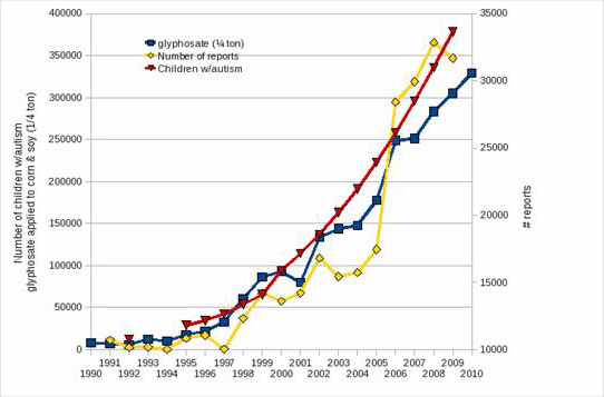
Dr. Seneff believes that when children are overexposed to glyphosate,
especially through consumption of the GM foods that are widely
prevalent in the American diet, they are more likely to react badly to
vaccination. To illustrate this point, Dr. Seneff and Nancy Swanson
plotted a graph showing autism trends in the US (as measured by autism
rates in the US school system), adverse vaccine reactions reported to
the VAERS system, and glyphosate application to GM corn and soy crops in
the US (Figure 3). As can be seen, the trends overlap almost entirely,
presenting “tantalizing links” between these variables. Dr. Seneff
infers from these findings that glyphosate is making vaccines far more
toxic than they would otherwise be.
Figure 3. Autism, glyphosate, and vaccine reactions in the US (Figure kindly provided by Nancy Swanson)
Summary
Taken together, the body of evidence elegantly assembled
by Dr. Seneff supports her hypothesis that the epidemic levels of autism
(and other diseases such as Alzheimer’s disease) currently seen in the
Western world are caused by a severe deficiency in sulfate supplies to
the brain. Under optimal circumstances, the pineal gland can synthesize
sulfate stimulated by sunlight and deliver it via melatonin sulfate to
the brain. However, aluminum, mercury, and glyphosate are working
synergistically to derail this process, and sunlight deficiency
(exacerbated by the misguided use of sunscreens containing aluminum
nanoparticles) is further contributing to the pathology.
About the Author
Claire Viadro, MPH, PhD, is a professional writer and
editor with two advanced degrees in public health. Her work has
included serving as past editor of Autism Science Digest magazine; co-editing Bugs, Bowels, and Behavior: The Groundbreaking Story of the Gut-Brain Connection; and authoring or coauthoring over 20 peer-reviewed publications primarily focused on women’s health.